Cellbytes – precision hematology with digital microscopy

The future is now

Cellbytes is the new standard in hematopathology. By integrating the entire cytomorphological analysis for both bone marrow and peripheral blood smears in one software, it’s easier than ever to modernize your diagnostic workflows.

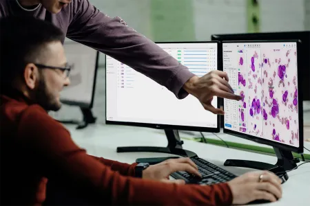
Two doctors analyzing the results of a slide using Cellbytes.

Objective and transparent

Cellbytes is a digital hematology system designed to augment hematopathologists, not replace them. Standardized, real-time digital assessment can enhance human performance and decrease subjectivity. Trained on millions of cells across diverse hematologic conditions, Cellbytes provides consistent, high-quality outputs. Every model prediction comes with clear visualizations and quantifiable data, ensuring transparency and supporting users to review what the software has identified.

Application details
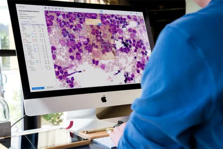
Doctor leaving a comment on an interesting slide region using Cellbytes.

Real-time analysis

Cellbytes is an automated digital morphology analyzer designed for high-throughput, parallelized batch analysis serving both small and large laboratories ensuring diagnostics will keep pace with your clinical need. The secure cloud-based computing is available wherever you are and 24/7. You can also retrieve historical slides in seconds to effortlessly track patient progression over time.

Analysis/evaluation workflow

Precision software optimized with industry-leading imaging

Out of all the supported scanners, the Evident SLIDEVIEW VS200 and Pramana HT Series are perfect matches for Cellbytes. Partnering with industry leading image quality and automated imaging enables you to focus on what matters most.

Our digital hematology platform also natively supports most major microscopes and scanners with output images in DZI, SVS, VSI, CZI, MRXS, DICOM, and OME-TIFF formats. Contact our team today to verify compatibility with your institution’s specific imaging system.

Close-up of a microscope scanning a hematopathological slide.

Flexible workflow integration

We built Cellbytes for healthcare professionals, the pharmaceutical industry, and academic researchers who need clearer insights from visual data. It combines the accuracy required for routine diagnostics, the versatility of consultation and collaboration, as well as the flexibility needed to discover new biological insights. Especially clinical workflow may not always be the most robust, but with our remote hematology review software, collaboration both within and out of your facility becomes easier.

Clinical workflow

Safe and secure

We are committed to data security. By strictly adhering to international standards and data privacy regulations (HIPAA, GDPR), we can guarantee users to retain complete control over their data.

Security

Full AI-assisted morphology analysis

Hematopathology is not just about counting cells. Cellbytes performs end-to-end cytomorphological assessment covering cellularity, sample adequacy, megakaryocyte counts, cell classification, and dysplasia detection. Results are summarized in a standardized report that can be seamlessly integrated into your laboratory information system.

A laptop on a desk showing a cell gallery in Cellbytes.

From cell images to clinical decisions

An automatic cell differential is one thing. But a software to support every step of cytomorphological assessment is another. Cellbytes is there to elevate your diagnostic work and create new opportunities from remote work, consultation and even collaboration across clinical laboratories.

Clinical use-cases

Quantitative morphometry

Cellbytes was created by researchers for researchers. Analyzing cell morphology requires strong domain knowledge spanning both hematology and hematopathology. Cellbytes democratizes this process by providing researchers and pharmaceutical teams with a certified platform to turn high-dimensional single-cell morphology into disease biomarkers and accelerate drug discovery.

Research use-cases

Latest news

3D render image of a bunch of small green and teal pill shapes hanging out in clusters.

Morphology Reveals CML Outcomes

Two peer-reviewed studies using the Cellbytes platform show how bone marrow morphology predicts CML treatment response and eligibility for treatment-free remission.

All news

Frequently asked questions

How can I use Cellbytes?

Cellbytes can be used by uploading high-resolution images of bone marrow aspirates or peripheral blood smears to the platform. The application analyzes these images using advanced machine learning algorithms, delivering quantitative results and visual annotations. It is accessible via a secure web interface—no installation required.

Can I test Cellbytes?

Yes, Cellbytes offers a trial version for institutions and professionals interested in evaluating its capabilities. You can request trial access by contacting the support team through the website.

How robust are the algorithms?

Cellbytes’s algorithms have been trained on a comprehensive real-world dataset comprising different pathologies and over 60,000 samples. The system achieves high accuracy in detecting and classifying various blood cell types and their abnormalities, with performance metrics benchmarked against expert diagnoses.

Will Cellbytes replace hematopathologists?

No, Cellbytes is designed to assist hematopathologists. It serves as a diagnostic aid that enhances efficiency and consistency. Final clinical decisions should always be made by a qualified medical professional.

Why should I use Cellbytes?

Cellbytes accelerates and enhances hematological analysis by providing AI-assisted interpretation of bone marrow and peripheral blood cell images. Cellbytes may facilitate remote working and consultation but also reduce diagnostic turnaround time and improve standardization, particularly in high-volume settings.

What type of images are supported?

Cellbytes supports high-resolution digital images of May-Grünwald Giemsa stained bone marrow aspirate and peripheral blood smear slides. Whole-slide images should be captured at 10-40x magnification and region-of-interest images should be acquired at 40-100x magnification. Cellbytes supports multiple imaging formats, such as DZI, SVS, VSI, CZI, MRXS, DICOM, and OME-TIFF.

Why is Cellbytes only cloud-based?

Being cloud-based ensures scalable computing resources and consistent algorithm performance across users. Software and algorithm updates can be deployed without delays. It also allows for secure data storage and easy collaboration between institutes, laboratories and specialists across geographic locations.

What is the price of the Cellbytes license?

Pricing depends on your usage volume. Contact the support team to test a trial version and discuss your needs. For research use, see the pricing models for research.

Can I use the application for research?

Absolutely. Cellbytes is increasingly used in research settings to study hematologic disorders, develop new biomarkers, and support clinical trials. It offers data export and integration with other medical data domains. Read more about how to use Cellbytes for research cases and what research has already utilized Cellbytes.

In research partnership with

Ready to digitize your cell morphology analysis?

Contact us to explore how Cellbytes can enhance your daily workflow.