100 times magnified image of blood cells, with annotated bounding boxes around white blood cells labeled by Cellbytes.

A new era

Digital cytopathology streamlines workflows by fostering synergy between physicians and technology. With AI managing time-consuming, quantifiable tasks, human experts apply their clinical judgment to verify and interpret results. To support interpretation, Cellbytes is the first platform to also provide reference values for healthy subjects. Remote collaboration becomes effortless, enabling seamless sample evaluation and colleague consultations. Cellbytes brings digital cytopathology to life.

Cellbytes workflow

  1. A doctor skimming through a drawer filled with bone marrow slides.

    Scan Slides

    Capture cytomorphological slides using your microscope.

  2. A rack of servers with networking cables running from one to another.

    Automatic Upload

    Images are securely uploaded to the cloud-based file storage.

  3. A screenshot of Cellbytes showing slide texture overlay mask.

    Feature Extraction

    Cellbytes analyzes key features from whole-slide and single-cell images.

  4. A screenshot of Cellbytes showing a list of cells with their labels.

    Physician Review

    Pre-classification results are reviewed and verified by a physician.

  5. A doctor reviewing a report, holding a pen in hand.

    Seamless Reporting

    Share a standardized digital report directly to your laboratory information system.

Key features

Sample triage

Prioritize urgent samples with a multi-parameter risk model.

Comprehensive image view

Access whole-slide images, numerical analysis, and visual masks.

Single-cell analysis

View single-cell images with classifications and potential dysplasia indicators.

Reference comparisons

Compare results with population-scale reference ranges.

Standardized reporting

Assess and share clinically essential findings in a structured report.

Collaborative sharing

Easily share slides and cell images with colleagues for consultations and meetings.

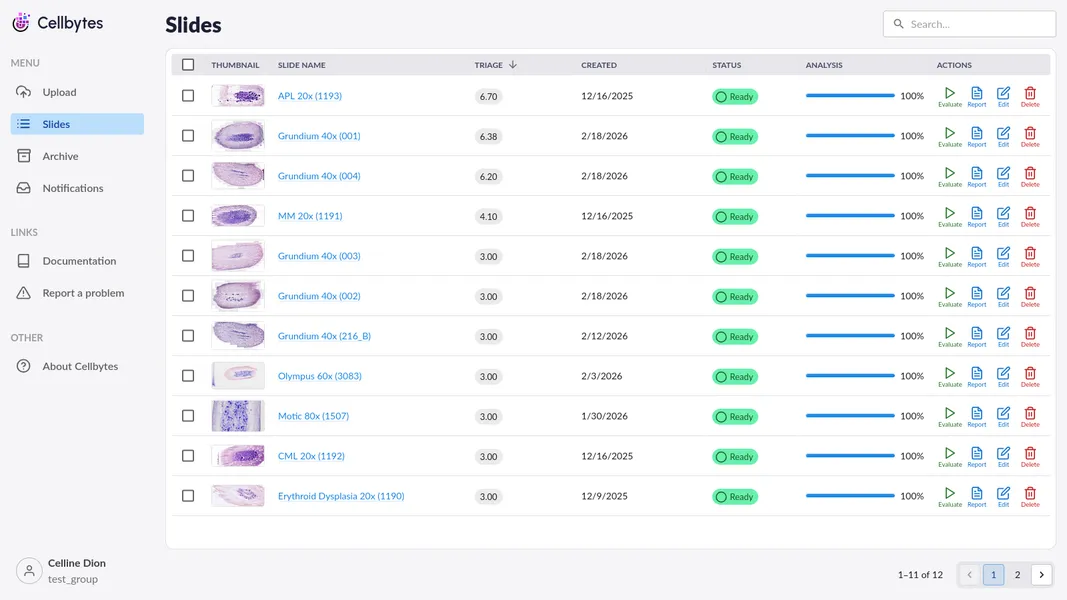
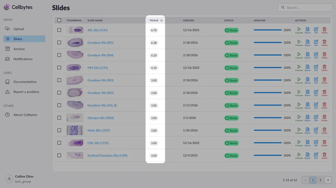
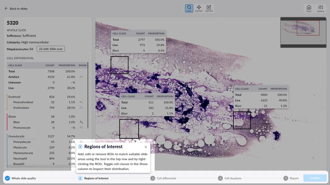
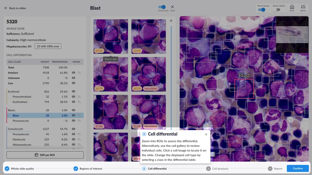
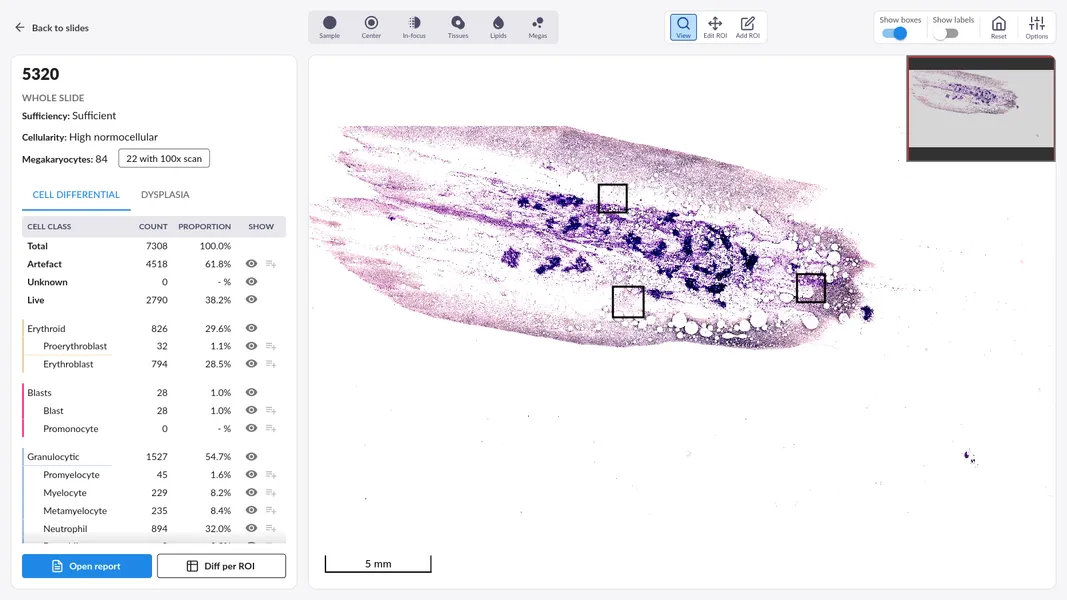
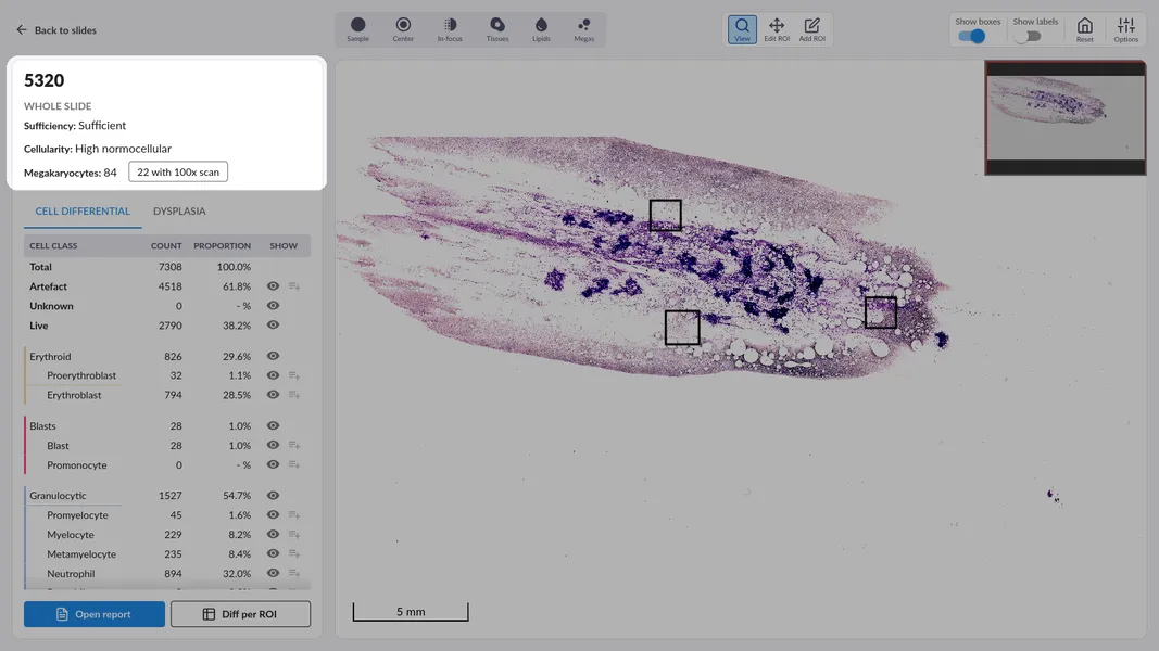
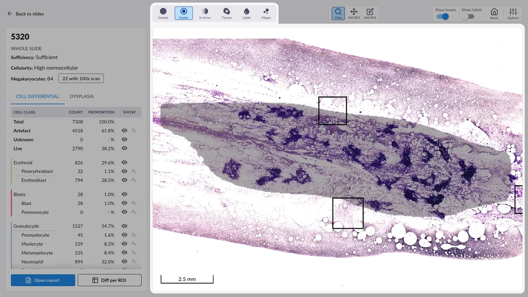
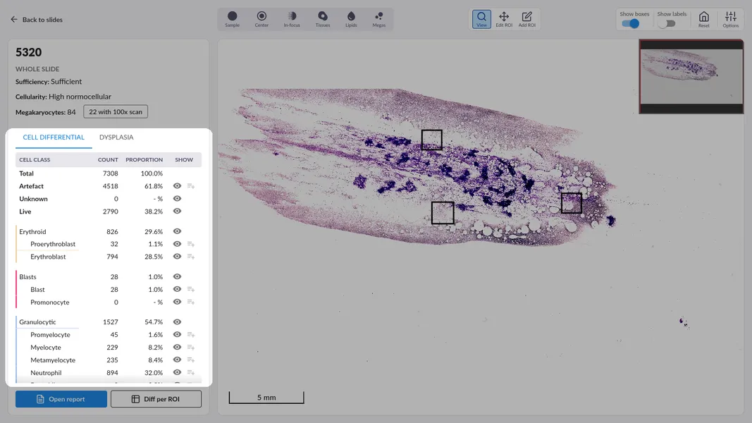
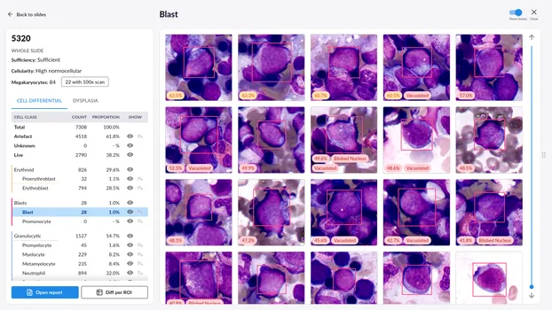
Comprehensive AI-powered analysis

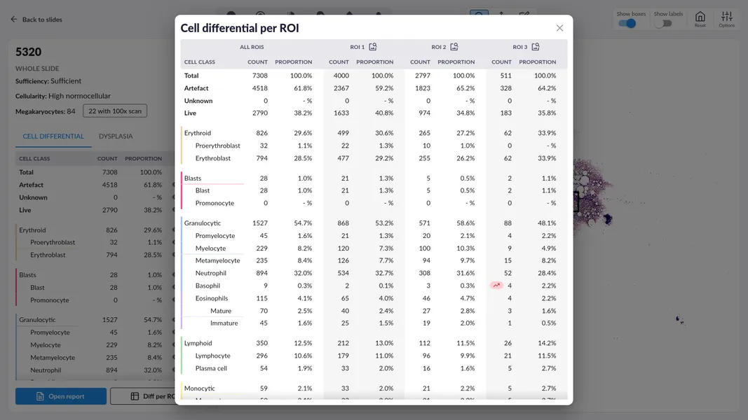
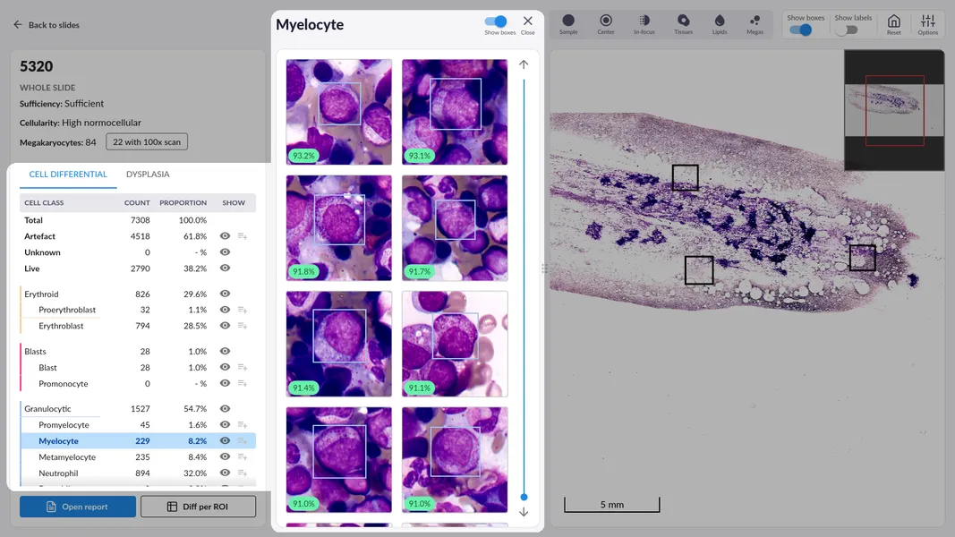
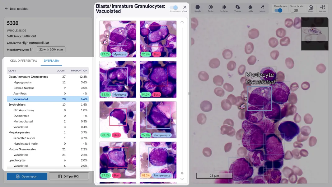
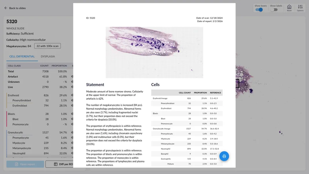
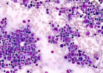
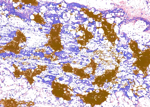
Our AI aims to perform nearly all the same diagnostic tasks as doctors, enhancing every step of the analysis process rather than just optimizing isolated aspects. By combining cell classification with morphology, dysplasia assessment, and sample quality analysis, we provide a fresh angle for research.

Intelligent triage

Our system ranks bone marrow samples by clinical urgency. By scoring cytomorphological features automatically, we ensure high-priority cases receive immediate attention.

Guided diagnostic workflow

Our system facilitates rapid adoption through step-by-step instructions and an intuitive introduction to the interface.

Guided diagnostic workflow

The evaluation flow will guide you through a sequence of analyses covering whole-sample analysis, selection of regions of interest, cell differential, dysplasia assessment and finally approving a standardized sample report.

Advanced interface for expert control

For experienced users, the advanced interface provides access to the full sample, offering total flexibility to conduct deep-dive analyses tailored to each case.

Comprehensive sample assessment

The system evaluates sample sufficiency, cellularity estimates to assess the density of hematopoietic cells and megakaryoycyte density, which are integral part of a comprehensive cytomorphological analysis.

Visual verification

Validate preclassification results with precision using diagnostic masks. Overlays highlight the exact regions factored into the analysis, providing full transparency into the system’s identification process.

Innovative cell differential

Cellbytes combines a powerful in-house foundation model trained with millions of cells and medical context to preclassify every cell identified within the sample. For targeted analysis, users can define specific regions of interest to maximize clinical accuracy.

Per-ROI detailed view

For a deeper dive, Cellbytes provides a detailed breakdown of results on a region-by-region basis to precisely compare different areas within the sample.

Review and refine automated classifications with ease. Our organized cell-by-cell list allows users to validate the system’s performance and maintain full diagnostic control by updating labels on the fly. Cellbytes ensures rapid correction and validation using an intelligent cell similarity measure.

Lineage-specific dysplasia analysis

A first in automated cytomorphology: detect and characterize dysplastic cells across multiple lineages rapidly and securely.

Seamless reporting

Finalize your evaluation with a single click. Approve the generated results and sign off on the sample to complete the digital workflow and archive the report.

Frequently asked questions

How can I use Cellbytes?

Cellbytes can be used by uploading high-resolution images of bone marrow aspirates to the platform. The application analyzes these images using advanced machine learning algorithms, delivering quantitative results and visual annotations. It is accessible via a secure web interface—no installation required.

Can I test Cellbytes?

Yes, Cellbytes offers a trial version for institutions and professionals interested in evaluating its capabilities. You can request trial access by contacting the support team through the website.

How robust are the algorithms?

Cellbytes's algorithms have been trained on a comprehensive real-world dataset comprising different pathologies and over 60,000 samples. The system achieves high accuracy in detecting and classifying various blood cell types and their abnormalities, with performance metrics benchmarked against expert diagnoses.

Will Cellbytes replace hematopathologists?

No, Cellbytes is designed to assist hematopathologists. It serves as a diagnostic aid that enhances efficiency and consistency. Final clinical decisions should always be made by a qualified medical professional.

Why should I use Cellbytes?

Cellbytes accelerates and enhances hematological analysis by providing AI-assisted interpretation of bone marrow cell images. Cellbytes will facilitate remote working and consultation but also reduce diagnostic turnaround time and improve standardization, particularly in high-volume settings.

What types of images are supported?

Cellbytes supports high-resolution digital images of May-Grünwald-Giemsa–stained bone marrow aspirate slides. Whole-slide images must be captured at 10x magnification and saved in an open-source format such as MIRAX. Region-of-interest images should be acquired at 100x magnification and saved in JPEG or MIRAX format.

Why is Cellbytes only cloud-based?

Being cloud-based ensures seamless updates, scalable computing resources, and consistent algorithm performance across users. It also allows for secure data storage and easy collaboration between institutes, laboratories and specialists across geographic locations.

What is the price of the Cellbytes license?

Pricing depends on your usage volume. Contact the support team to test a trial version and discuss your needs.

Can I use the application for research?

Absolutely. Cellbytes is increasingly used in research settings to study hematologic disorders, develop new biomarkers, and support clinical trials. It offers data export and integration with other medical data domains.